In this article, we shall study blood vessels and their types: arteries, veins, and capillaries. Study of blood vessels is called Angiology.

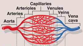
Blood vessels form a network of blood circulating vessels starting from the heart (left ventricle) arteries arterioles capillaries venules veins back to the heart (right auricle).
Arteries and Arterioles:

Arteries carry blood from the heart to the capillaries of the organs in the body.
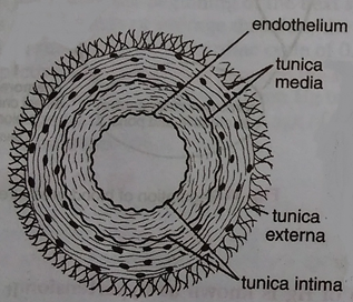
The walls of arteries are thicker than those of veins. The smooth muscle and elastic fibres that make up their walls enable them to withstand the high pressure of blood as it is pumped from the heart. Histologically the wall of the artery is made up of three layers namely, outer tunica externa, middle tunica media and inner tunica interna. The artery is characterised by the presence of thick and muscular tunica media.
Each artery expands when the pulse of blood passes through and the elastic recoil of the fibres cause it to spring back afterwards, thus helping the blood along. This is known as secondary circulation, and it reduces the load on the heart.
Other than the pulmonary arteries, all arteries carry oxygenated blood. The aorta carries oxygenated blood from the left ventricle to all parts of the body except the lungs. It has the largest diameter (25mm) and carries blood at the highest pressure.
As the aorta travels away from the heart, it branches into smaller arteries so that all parts of the body are supplied. The smallest of these are called arterioles. Arterioles can dilate or constrict to alter their diameter and so alter the flow of blood through the organ supplied by that arteriole.
Two organs which always have the same blood flow are the brain and the kidneys. Main organs to have blood flow reduced are the guts (between meals), muscles (when resting) and skin (when cold).
Capillaries:

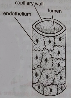
Arterioles branch into networks of very small blood vessels, the capillaries. These have a very large surface area and thin walls that are only one (epithelial) cell thick.
It is in the capillaries that exchanges take place between the blood and the tissues of the body. Capillaries are also narrow. This slows the blood down allowing time for diffusion to take occur. In most capillaries, blood cells must flow in single file. Tissue fluid is formed in the capillaries, for their walls are leaky
Venules and Veins:

After leaving the capillaries, the blood enters a network of small venules, which feed into veins. These, in turn, carry the blood back to the atria of the heart.
Like arteries, the walls of veins are lined with epithelium and contain smooth muscle. The walls of veins are thinner and less elastic than arteries, but they are also more flexible. Histologically the wall of vein also made up of three layers namely, outer tunica externa, middle tunica media and inner tunica interna. But all the layers are thin.
Veins tend to run between the muscle blocks of the body and nearer to the surface than arteries.
The larger veins contain valves that maintain the direction of blood flow. This is important where blood must flow against the force of gravity.
The flow of blood in veins is helped by contractions of the skeletal muscles, especially those in the arms and legs. When muscles contract they squeeze against the veins and help to force the blood back towards the heart. Once again, this is known as secondary circulation.