Science > Biology > Zoology > Histology > Epithelium or Epithelial Tissue
In this article, we shall study about the epithelium or epithelial tissue.
Higher-level organisms have multicellular structure. The cells differ from one another in their shape, size. Cells arise from pre-existing cells by the process of cell division.
In multicellular organisms, cells are broadly classified as somatic cells and germ cells. Somatic cells mean body cells and present all over the body. They are responsible for all body-related activities. Germ cells are associated with reproduction and present in organs of reproduction. Cell work in groups called tissue. A group of cells having the same embryonic origin, structure, and function is called tissue. Tissues combine together to form large functional units called organs. A number of organs work in coordination and forms an organ system.
Somatic cells are grouped into four types of somatic tissues epithelial, connective, muscular and nervous.
Epithelial Tissue or Epithelium (Greek epi- upon; thelio – grows):
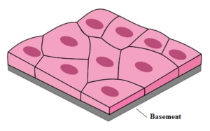
The term epithelium was introduced by a Dutch anatomist Ruysch, which means the tissues that grow upon other tissues. Epithelial tissues line body surfaces, hollow organs, blood vessels, ducts, and cavities, as well as form glands. They have two distinct body surfaces inner and outer. The apical surface is exposed to the body cavity or exterior, while the basal surface is adjacent to the underlying connective tissue.
Epithelial linings of some hollow organs or cavities are moist due to the secretion of mucus by the epithelial tissue. Such lining of epithelial cells is called a mucous membrane or mucosa. Epithelia contain no blood vessels (they are non-vascular) and are dependent upon the underlying connective tissue for nutrients. They have their own nerve supply. Cells are placed on a thin, double-layered, noncellular basement membrane.
They have a capacity for self-renewal and repair. The old injured cells are shaded off regularly. The function of epithelial tissue varies depending on its location.
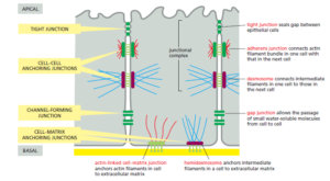
Junctions Between Two Epithelial Cells:
The cells of the tissue are closely placed and connected to each other via cellular junctions. Very little or no intracellular space present. The cells are held together by intercellular junctions such as desmosomes, interdigitations, tight junctions, gap junctions, intercellular bridges and closed fitted folds.
The tight junction stops substances from leaking across a tissue. Adhering junction keeps the two neighbouring cells cemented together. Gap junction helps in communication between the two cells by connecting the cytoplasm of the two cells for easy transfer of ions and small molecules.
Functions of Epithelial Tissues:
- The epithelium of the skin protects the underlying tissues from mechanical damage, ultraviolet light, dehydration and invasion by bacteria
- The columnar epithelium of the intestine secretes digestive enzymes into the intestine and absorbs the products of digestion from it.
- An epithelium also lines our respiratory tract. It secretes mucus which keeps it from drying out and traps inhaled dust particles. Most of its cells have cilia on their apical surface that propel the mucus with its load of foreign matter back up to the throat.
| Function | Description |
| Protection | Protects underlying tissues from mechanical and chemical injury, from dehydration and from the entry of germs. |
| Secretion | Gladular epithelium secretes mucus, and important substances required for metabolism like gastric juices, hormones etc. |
| Absorption | Epithelial lining of micro villi of small intestine absorb digested food material. |
| Excretion | lining of uriniferous tubules of kidney does the function of excretion of nitrogenous waste. |
| Reproduction | Epithelium of seminiferous tubules in males and ovaries in female produce gametes. |
| Respiration | lining of alveoli in lung provides the surface for gaseous exchange. |
| Conduction | In respiratory tract ciliated epithelium moves mucus. In fallopian tubes ovum is moved forward towards uterus by ciliated epithelium. |
| Sensation | Sensory epithelium in sense organs sends nerve impulses from the environment to nervous system |
| Exoskeleton Formation | Exaskeletol structures like nails, scales, hairs, horns, feathers, hoofs are formed from surface epithelium. |
Types of Epithelial Tissues:
Simple Epithelium:
It is made up of a single layer of cells which is useful in diffusion, osmosis, filtration, secretion, and absorption. The cells rest on the basement membrane. A stratified epithelium consists of two or more layers of cells. The lowermost layer rests on the basement membrane. The function of simple epithelial tissue varies depending on its location. Simple epithelium can be further classified as squamous, cuboidal, columnar, ciliated and glandular epithelium.
Simple Squamous Epithelium:
It is present in the peritoneum of coelom and endothelium the inner lining of blood vessels. The inner lining of blood and lymph vessels provides a smooth surface that reduces friction as blood travels through the vessels.
They are also found in alveoli and capillaries of lungs where gas exchange occurs, kidney glomerulus and tubules where filtration and diffusion processes form urine, capillaries where diffusion and osmosis occur.
The cells are polygonal in shape. thin, delicate and flat. The nucleus is present at the centre. As they look like layered tiles from above, they are called pavement epithelium. Their main function is the filtration and diffusion of material.
Simple Cuboidal Epithelium:

It is found in thyroid glands and kidney. They are also found in exocrine glands (lining of salivary and pancreatic ducts) and endocrine glands, ducts of endocrine glands. Due to larger cytoplasmic volumes, they can perform complex functions like absorption and secretion.
The cells are cube-shaped with a central round nucleus. Their primary function is secretion, excretion, and absorption. When performing absorption function they bear microvilli on their free ends. it increases the area of absorption. This gives a brush-like appearance to their apical end.
Simple Columnar Epithelium:
Nonciliated columnar epithelium forms the inner lining of the stomach, intestine, internal organs and ducts of exocrine glands. The ciliated columnar epithelium is present in the respiratory tract and in the fallopian tubes of the female reproductive tract. The primary function of these cells is the absorption of nutrients. With the largest cytoplasmic volumes of all epithelia, these cells possess the organelle density and energy reserves to engage in the most complex and efficient secretory or absorptive functions.
The cells are tall, pillar-like and broader t the free end. They appear rectangular in vertical section and polygonal in surface view. The basal end is fixed to the basement membrane. The nucleus is oval and located at the basal end. They are further classified as ciliated columnar epithelium and nonciliated columnar epithelium.
Ciliated Columnar Epithelium:
Ciliated columnar epithelia are found in small bronchioles of the respiratory tract and in the fallopian tubes of the female reproductive tract. In the respiratory tract the cilia aid in the movement of mucus and foreign particles. In the fallopian tubes, cilia move reproductive cells (ovum) towards the uterus.
Non-Ciliated Columnar Epithelium:
Nonciliated columnar epithelia with microvilli line the small intestine where 90% of absorption from the digestive tract occurs. It increases the absorption area of the intestine.In the intestine many of these cells secrete mucus which gets accumulated at the apical end of the cells. These cells are called goblet cells.
The unicellular gland or goblet cell is a specialized columnar cell of mucous membranes that secrete mucus for protection
In pseudostratified epithelium, nuclei are at several different levels, giving an appearance of an epithelium composed of layers of cells (a stratified epithelium). However, all of the cells of a pseudostratified epithelium are in contact with the basement membrane. The term pseudo (false) indicates that this is not a stratified epithelium. In most locations in the body pseudostratified epithelia are ciliated. The lining of the trachea is an example of the ciliated pseudostratified epithelium.
Glandular Epithelium:
These are the specialized group of epithelial cells capable of synthesizing substances like enzymes, hormones, sweat, oil, mucus, etc. Their secretions are carried into ducts on the surface or into the blood. The structure formed by such glandular epithelium is called gland. Glands are further classified as endocrine glands and exocrine glands.
Endocrine Glands:
Endocrine glands pour their secretion directly into the bloodstream hence they are also called ductless glands. These are the glands which lose all contact with the epithelial surface from which they develop. They pour their secretion directly into the bloodstream and is circulated throughout the body and is utilized by specific target tissues. Some examples of endocrine glands are pituitary, thyroid, parathyroid, ovary, testis, adrenal, Isles of Langerhans.
Exocrine Glands:
Exocrine glands have their own ducts through which their secretion is carried out to the site of action. These glands have an extensive blood supply and they are under nervous and hormonal control. Depending on the mode of secretion these glands are classified into three types.
- Holocrine Glands: The entire cell disintegrates after secretion. e.g. sebaceous glands
- Merocrine Glands: Secretion is thrown out of the cells by the process of diffusion. The cell remains intact.g. sweat, salivary, intestinal glands.
- Apocrine Glands: Apical parts of cells are shed off to discharge secretion. e.g. mammary glands.
Some examples of exocrine glands are salivary glands, tear glands, gastric and intestinal glands.
Compound Epithelium:
They consist of more than one layer of cells. The innermost layer of cells rests on the basement membrane. Due to the multilayer structure, they do not perform the function of secretion and absorption. Their function is to protect inner tissues from mechanical, chemical, thermal or osmotic attacks.
The compound epithelium is classified into two types a) Stratified squamous epithelium and b) Transitional epithelium.