Science > Biology > Botany > Reproduction in Plants > Androecium and Gynoecium
The flower is the reproductive unit in the angiosperms. It is meant for sexual reproduction. A typical flower has four different kinds of whorls arranged successively on the stalk or pedicel, called thalamus or receptacle. These whorls are calyx, corolla, androecium and gynoecium. Calyx and corolla are accessory organs, Androecium and gynoecium are reproductive organs. When a flower has both androecium and gynoecium, it is bisexual. A flower having either only stamens or only carpels is unisexual.

Parts of Flower involved in sexual reproduction:
Androecium:

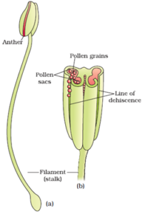
The androecium is composed of stamens. Each stamen which represents the male reproductive organ consists of a stalk or a filament and an anther. Each anther is usually bilobed and each lobe has two chambers, the pollen-sacs. The microspores (pollen grains) are produced in pollen-sacs. A sterile stamen is called staminode.
Gynoecium:

The gynoecium is the female reproductive part of the flower and is made up of one or more carpels. A carpel consists of three parts namely stigma, style and ovary. The ovary is the enlarged basal part, on which lies the elongated tube, the style. The style connects the ovary to the stigma. The stigma is usually at the tip of the style and is the receptive surface for pollen grains. Each ovary bears one or more ovules attached to a flattened, cushion-like placenta. Ovules develop into seeds and the ovary matures into a fruit.
Structure of Anther (Microsporangium):
In a transverse section, a typical microsporangium appears near circular in outline. It is generally surrounded by four wall layers the epidermis, endothecium, middle layers and the tapetum.
The epidermis consists of flattened cells and is protective in the function. It is outermost common wall layer of the anther. Endothecium is internal to the epidermis. Cells of endothecium help for dehiscence of anther at maturity. Middle layers are internal to endothelium. They are of three layers of parenchyma cells. The cells of these layers degenerate on maturity and the two pollen sacs of each lobe merge to form a single chamber. Tapetum surrounds sporanenous cells and provide nutrition to developing pollen grains.
As each cell of the sporogenous tissue is capable of giving rise to a microspore tetrad. Each one is a potential pollen or microspore mother cell (PMC). The process of formation of microspores from a pollen mother cell through meiosis is called microsporogenesis. The microspores, as they are formed, are arranged in a cluster of four cells–the microspore tetrad. As the anthers mature and dehydrate, the microspores dissociate from each other and develop into pollen grains.
Inside each microsporangium, several thousands of microspores or pollen grains are formed that are released with the dehiscence of the anther
Structure of pollen grains:
The pollen grains represent the male gametophytes. It has a prominent two-layered wall. The hard outer layer called the exine is made up of sporopollenin which is one of the most resistant organic material known. It can withstand high temperatures and strong acids and alkali. No enzyme that degrades sporopollenin is so far known.
Pollen grain exine has prominent apertures called germ pores where sporopollenin is absent. The inner wall of the pollen grain is called the intine. It is a thin and continuous layer made up of cellulose and pectin. The cytoplasm of the pollen grain is surrounded by a plasma membrane.
When the pollen grain is mature it contains two cells, the vegetative cell and generative cell. The vegetative cell is bigger, has abundant food reserve and a large irregularly shaped nucleus. The generative cell is small and floats in the cytoplasm of the vegetative cell. It is spindle-shaped with dense cytoplasm and a nucleus.
Development of Male gametophyte:
Before pollination in pollen sack:
The protoplast of pollen grain divides by mitosis to form to unequal cells. The smaller cell is called the generative cell. It has a large nucleus, thin cytoplasm and it lacks reserve food and vacuole. The large Cell is called vegetative cell or tube cell. It has large vacuole, cytoplasm, nucleus and reserve food.
After pollination on the stigma:
When the two-celled pollen grain comes in contact with sugary stigmatic secretion it absorbs it. Due to absorption of secretion, the pressure of cytoplasm on the intine increases and the intine of pollen grain comes out of germ pore to form a pollen tube. The pollen tube starts growing towards the ovule trough style due to chemical stimulus in the ovary.
The tube nucleus, cytoplasm and generative cell starts migrating into the pollen tube. The generative cell divides by mitosis to form two haploid non-motile male gametes.
Gynoecium:
The gynoecium represents the female reproductive part of the flower. The gynoecium may consist of a single pistil (monocarpellary) or may have more than one pistil (multicarpellary). Each pistil has three parts the stigma, style and ovary. The stigma serves as a landing platform for pollen grains. The style is the elongated slender part beneath the stigma.
The basal bulged part of the pistil is the ovary. Inside the ovary is the ovarian cavity (locule). The placenta is located inside the ovarian cavity. Arising from the placenta are the megasporangia, commonly called ovules. The number of ovules in an ovary may be one (wheat, paddy, mango) to many (papaya, water melon, orchids).
Structure of Ovule:
The ovule is a small structure attached to the placenta by means of a stalk called funicle. The body of the ovule fuses with funicle in the region called hilum. Thus, hilum represents the junction between ovule and funicle. Each ovule has one or two protective envelopes called integuments. Integuments encircle the ovule except at the tip where a small opening called the micropyle is present. Opposite the micropylar end, is the chalaza, representing the basal part of the ovule.
Enclosed within the integuments is a mass of cells called the nucellus. It consists of many diploid parenchyma cells. Cells of the nucellus have abundant reserve food materials. Located in the nucellus is the embryo sac or female gametophyte. An ovule generally has a single embryo sac formed from a megaspore through reduction division.
Functions of Parts of Ovule:
- Funicle: To support, projection and conduction
- Nucellus: Development of female gametophyte takes place in it.
- Integuments: Protection to nucellus and embryo sac
- Micropyle: It forms a passage for pollen tube to enter in the ovule.
- Antipodals: They are accessory cells and degenerate after fertilization.
The process of formation of megaspores from the megaspore mother cell is called megasporogenesis.
Ovules generally differentiate a single megaspore mother cell (MMC) in the micropylar region. The diploid MMC (2n) undergoes meiosis to form a tetrad of haploid megaspores (n)
Development of Female Gamete:
The chalazal megaspore remains functional, while the other three degenerate gradually. The functional megaspore undergoes enlargement and develops into the female gametophyte. The nucleus of the functional haploid megaspore divides mitotically to form two nuclei which move to the opposite poles, forming the 2-nucleate embryo sac. Two more sequential mitotic nuclear divisions result in the formation of the 4-nucleate and later the 8-nucleate stages of the embryo sac.
There is cellular organization in which 3 celled egg apparatus is formed at mycropylar end and constitute the egg apparatus. The egg apparatus, in turn, consists of two synergids and one egg cell. Three cells are at the chalazal end and are called the antipodals. The large central cell, has two polar nuclei. Thus, a typical angiosperm contains embryo sac, at maturity, though 8-nucleate is 7-celled.
One reply on “Androecium and Gynoecium”
This gave me more than enough information to make sense of the flower structure lecture we had yesterday as part of my horticultural course. This information was well written and made sense of what yesterday appeared confusing, thanks,