Since the heart relies on aerobic respiration to supply its energy needs, cardiac muscle cells are richly supplied with mitochondria.
Our hearts beat about once every second of every day of our lives or over 2.5 million times in an average life span. The only time the heart gets a rest in between beats.
Position:
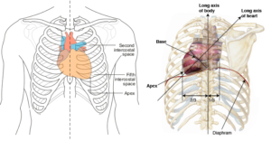
It is a first—sized cone-shaped hollow muscular organ that is situated between the two lungs and above the diaphragm and behind the sternum in the chest. The base is tilted to the left. It is guarded by the rib cage. The heart contracts at regular intervals, forcing blood through the circulatory system.
A thin, tough membrane called the pericardium surrounds and holds the heart in position. This membrane has a slippery surface inside and allows the heart to move easily when it beats. The fluid present between the pericardial membranes is called the pericardial fluid. It helps to reduce friction between the walls of the heart and surrounding tissues when the heart is beating.
External Structure

Posterior View

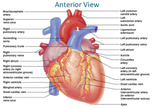
Structure of Heart
It is a hollow, muscular organ that contracts at regular intervals, forcing blood through the circulatory system. It is cone-shaped, about the size of a fist, and is located in the centre of the thorax, between the lungs, directly behind the sternum (breastbone). The base is tilted to the left.
Pericardium (heart enclosure):
It is made up of two layers parietal layer (outer layer) and visceral layer attached to the heart. Between these two layers pericardial fluid is present, which acts as a shock absorber. It has two layers.
- Fibrous pericardium: It is tough, inelastic and fibrous connective tissue.
- Serous pericardium: Its function is the protection of the heart.
Heart wall:
It has three layers.
- Epicardium: It is made up of single layer flat epithelial cells called the mesothelium.
- Myocardium: It is made up of cardiac muscles responsible for the beating of the heart
- Endocardium: It is made up of single layer flat epithelial cells called the endothelium.
Cardiac Muscles:
The cardiac muscles are not under the conscious control of the nervous system and can generate its own electrical rhythm (myogenic). For the same reasons, cardiac muscle cannot respire anaerobically and so the muscle cannot get tired (or develop cramp!). Cardiac muscle has a rich supply of blood, which ensures that it gets plenty of oxygen. This is brought to the heart through the coronary artery.
The heart is divided into two halves the right side and the left side by a thick, muscular wall called the atrioventricular septum. Each half has two chambers the upper auricles (singular atrium) and the lower ventricles. The walls of auricles are comparatively thinner, while walls of ventricles are comparatively thicker, muscular walls. Thus the heart has four chambers. The right side of the heart contains deoxygenated blood while the left side of the heart contains oxygenated blood.
The right ventricle pumps blood to the lungs through pulmonary arteries. The right auricle receives blood from the rest of the body through superior vena cava and inferior vena cava. The superior vena cava returns blood from the head and arms; the inferior vena cava from the rest of the body (except the lungs!). The entry of blood from vena cavae is guarded by valves. During joint diastole blood enters from vena cavae into the right auricle.
The entry of blood from the right auricle to the right ventricle is guarded by an atrioventricular valve having three flaps and called the tricuspid valve.
During the atrial systole auricles contract, the tricuspid valve opens and the blood from the right auricle is emptied in the right ventricle.
The opening of the right ventricle in pulmonary arteries is guarded by the pulmonary valve. During ventricular systole the tricuspid wall is closed, the ventricles contract, the pulmonary valve opens. The blood from the right ventricle is forced into pulmonary arteries. Pulmonary arteries are further bifurcated into two vessels carrying blood to the left and right lungs.
This deoxygenated blood gets oxygenated in the lungs and returns back to the left auricle of the heart through four pulmonary veins (two from each lung). During joint diastole, the left auricle is filled with blood coming from the lungs.
The flow of blood from the left auricle to the left ventricle is guarded by a valve having two flaps called a bicuspid valve. During auricular systole the left auricle contracts, the bicuspid or mitral valve opens. Blood is forced into the left ventricle.
During ventricular systole the left ventricle contracts, the blood in it is forced at very high pressure through another semi-lunar valve (the aortic valve), into the aorta. Aorta carries blood throughout the body.
After moving through different parts of the body, the blood return backs to right auricle of heart through vena cavae.
Diastole:
When all the heart muscles relax, deoxygenated blood from the head and the body enters the right auricle through the superior vena cava and the inferior vena cava respectively. At the same time, oxygenated blood from the lungs enters the left auricle through the pulmonary vein. This phase is known as diastole.
Systole:
When auricles and ventricles contract the state is called systole. There are two systoles. a) auricular systole and b) ventricular systole. During auricular systole, the auricles filled with blood, contract, forcing the blood into the ventricles. During ventricular systole, the right ventricle contracts and forces deoxygenated blood to lungs through pulmonary arteries and left ventricle contracts and force oxygenated blood to different parts of the body through the aorta.
Physiologically heart can be myogenic or neurogenic
Myogenic Heart:
In this heart, the cardiac movement is initiated by cardiac muscles themselves. This type of heart is found in molluscus and vertebrates.
Neurogenic Heart:
The cardiac movement is initiated by the nerves arising from the brain. This type of heart is found in invertebrates except for moluscus.