
Science > Biology > Human Anatomy and Physiology > Cardiovascular System > The blood, an Overview
In the Cardiovascular system, the ‘‘heart’’ (cardi) pumps the blood in a ‘‘little circle’’ (circul), which travels through ‘‘little vessels’’ (vascul). In human, the transportation is done through blood circulatory system and lymphatic system. Thus two fluids move through the circulatory system: blood and lymph. The blood, heart, and blood vessels form the Cardiovascular System. The lymph, lymph nodes and lymph vessels form the Lymphatic System. Human blood circulatory system has three main components. A fluid (blood), tubing (arteries, veins and capillaries) and a pump (the heart).
Study of blood is called haematology. It is a fluid connective tissue. It is bright red, slightly alkaline, salty viscous fluid heavier than water.
The Constituent of the Blood:
In this article, we shall only take an overview of the composition. In the next article, each constituent is discussed in detail.
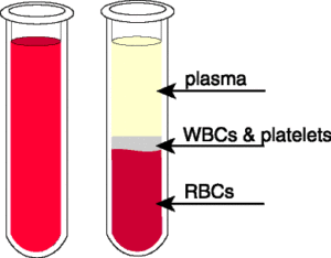
When a human blood sample is prevented from clotting and spun in a test tube (centrifuged), in a machine called a centrifuge, the blood separates into a straw coloured liquid called plasma and a dark brown mass of blood cells. The lower layer consists of white blood cells, blood platelets, and red blood cells. Collectively, these are the formed elements, which make up about 45% of the total volume of whole blood; the percentage of blood attributed to red blood cells is called the hematocrit. The hematocrit is defined as the percentage of blood volume that is occupied by erythrocytes. The normal hematocrit is approximately 45 percent in men and 42 percent in women.
The upper layer is plasma, which contains a variety of inorganic and organic molecules dissolved or suspended in water. Plasma accounts for about 55% of the total volume of whole blood.
Characteristics of Human blood:
Study of blood is called haematology. Blood is a fluid connective tissue. It is bright red, slightly alkaline (pH 7.3 to 7.5), salty viscous fluid heavier than water. the pH of blood is more in arteries than that in veins. The viscosity of blood is 5 to 6 times that of water. An adult has a blood volume of approximately 5.5 litres. It forms 6 to 10 % of the body weight. Blood is the only tissue that exists in both the liquid and solid state simultaneously.
Volume of Blood and Different Constituents:
The volume of blood in an average-sized person is approximately 5.5 L. Now hematocrit is 45 percent of the total volume,
Then, Erythrocyte volume = 0.45 x 5.5 L = 2.5 L
Since the volume occupied by the leukocytes and platelets is normally considered negligible, the plasma volume equals the difference between blood volume and erythrocyte volume; therefore, in our average person
Plasma volume = 5.5 L – 2.5 L = 3.0 L
Plasma:
The plasma consists of 90 to 92% water and 8% to 10% proteins, salts, hormones, enzymes, waste products and other various chemicals. Most of the solute part about 7% is proteins. These include antibodies that help to protect the body from diseases, fibrinogen that helps the blood to clot. The waste product includes urea and carbon dioxide. Hormones are the chemical messenger, which help to coordinate different body functions.
Plasma obtained from blood donation may be converted to a powdered form for storage. During the transfusion, it is dissolved in sterile distilled water and can be administered at once. This method saved the lives of many during World war.
Red Blood Cells (RBCs):
They are also called erythrocyte. They are produced inside the bone marrow. They have a lifespan of about 100 to 120 days after which they are destroyed by the liver. They are the most common type of blood cell (5.1 to 5.8 million per cubic mm). They are non-nucleated, small in size, round and biconvex in shape. They are able to fold and bend as they are forced through the smallest blood vessels.
The strong red colour of blood is due to the large number of RBCs. Red blood cells contain haemoglobin which gives them their red colour and enables them to carry oxygen from lungs to different parts of the body. They also carry carbon dioxide from different parts of the body to the lungs. Their main function is to carry oxygen from the lungs to the tissues.
White blood cells (WBCs):
They are also called leucocytes. Many white cells are made in the bone marrow. Their lifespan is 3 to 4 days. They are colourless and they have a nucleus. WBCs are larger than red cells but they are lesser in number. (About 5000-7000 per cubic millimetre of blood). They are further classified as lymphocytes and phagocytes. The nucleus of each type has a characteristic shape. When they travel in the blood they are more or less spherical, but they flatten and continuously change their shape along the inside walls of the blood vessels.
WBC’s rid the body of pathogens in the process called phagocytosis. In this process, the WBC surrounds, engulfs and “eats” the invading pathogen.
Platelets :
Platelets are also called thrombocytes. They are made in the bone marrow and have a lifespan of 8-14 days. They are much smaller than red cells. One cubic millimetre of blood contains about a quarter of million platelets. Their function is to help the blood clot. Clotting prevents loss of blood from wounds.
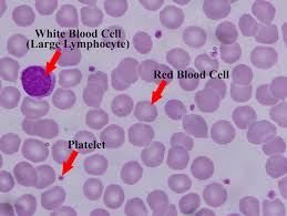
Observing Blood Under Microscope:
- Clean the skin of your finger with a swab of cotton dipped in ethanol. With a sterile needle prick your finger so that a drop of blood comes out.
- Place the drop of blood at one end of a microscope slide. With another slide spread the blood over the surface to form a smear. Let it dry and then examine it under a microscope. We can observe red blood corpuscles.
- Now cover the smear with Leishman’s stain and leave it for five minutes. Then wash the stain off with tap water gently. Let the slide dry and then examine it under the microscope again. Now, we can observe white blood corpuscles.
Functions of Human blood:
Transportation:
The blood moves from the heart to all the various organs, where exchange with tissues takes place across thin capillary walls. Blood collects oxygen from the lungs and nutrients from the digestive tract and transports these to the tissues. It delivers enzymes and chemical messengers to cells and tissues. Various organs and tissues secrete hormones into the blood, and blood transports these to other organs and tissues, where they serve as signals that influence cellular metabolism. It delivers water, vitamins and minerals to cells. It carries carbon dioxide from cells, tissues and carries it to lungs for disposal. It carries waste materials like urea and other chemical wastes and carries them to the liver and kidneys for disposal. It carries antibodies from place to place in the body. It carries vitamins and enzymes.
Protection:
The blood defends the body against invasion by pathogens (microscopic infectious agents, such as bacteria and viruses) in several ways. Certain blood cells are capable of engulfing and destroying pathogens, and others produce and secrete antibodies into the blood. White blood corpuscles (WBC) fight against disease-causing germs that harm the body.
It prevents the loss of blood after an injury by clotting. Blood helps in the repair process after a cut or other injury. Without blood clotting, we could
bleed to death even from a small cut.
Regulation:
Blood Picks up excess body heat and brings it to the skin to be excreted. The sweat is formed on the skin. Which is evaporated and heat required for it is taken from the body. Hence the body cools down. It controls the amount of water in the body. The salts and plasma proteins in blood act to keep the
liquid content of blood high. In this way, blood plays a role in helping to maintain its own water-salt balance. It also helps to regulate the amount of chemical substance in the tissues of the body. Due to the presence of buffers in the blood, it also helps to regulate body pH and keep it relatively constant.
Blood Donation:
Anybody who is healthy weighs over 50 kg and is between the age of 18 years and 65 years can donate blood. An adult has a blood volume of approximately 5 litres. A donor may give up to half a litre of blood at one time. This is quickly replaced by the body.
Many donors give blood regularly. It is immediately mixed with a chemical which prevents it from clotting and also provides food for the living cells. The blood is then stored in a refrigerator until it is required. Similarly, sodium citrate is added to it to avoid coagulation. The place where the blood is stored is called a blood bank.
Blood groups:
Human blood group is determined by the antigens present on the surface of RBC’s. Blood groups are inherited and do not change throughout life. Human blood is classified into 4 main groups: A, B, AB and O. Each can be either Rhesus + ve or Rhesus – ve, giving 8 groups in all. Blood grouping is the identification of the antigens in a blood sample. This system is called ABO system.
An individual’s RBC’s may carry an A antigen, a B antigen, both A and B antigens, or no antigen at all. These antigen patterns are called blood types A, B, AB and O, respectively. Type AB is known as a universal recipient, meaning that they can receive any type of blood, while O is the universal donor, meaning they can donate blood to anyone
Blood transfusion:
Blood transfusion is the transfer of blood that is taken from one person, into the bloodstream of another person. The person who gives blood is called the donor. The person who receives blood is called the recipient. When there is a loss of blood suddenly due to an accident, or because of the bursting of a blood vessel, there is a danger that not enough blood will be left to maintain the circulation. In such a case, the patient may lose consciousness due to low blood pressure and hence less supply of oxygen to tissues. Losing blood is called haemorrhage. To restore the blood volume and to provide more red cells, a blood transfusion is carried out.
Before doing blood transfusion the compatibility between the groups of the donor and the recipient should be checked. If the blood of the donor is not compatible with the blood of the patient, the red cells in the patient’s blood will stick together. This may lead to death.
Clotting of Blood:
Human blood contains heparin and antithrombins as anticoagulants, it prevents the blood to clot inside the blood vessels. As soon as blood vessel ruptures, bleeding starts. The conversion of liquid blood into semisolid jelly is called blood coagulation or blood clotting. Platelets adhere to the site of the wound and release clotting factors known as prothrombin. Prothrombin is inactive. At the site of rupture the platelets and injured tissues release thromboplastin which initiates the formation of enzyme prothrombinase. In the presence of Ca, ions prothrombinase converts inactive prothrombin to active thrombin. Thrombin converts soluble fibrinogen into fibrin. The fibrin forms a net to enmesh platelets blood cells and plasma to form a clot.
Previous Topic: Circulatory System and its Types
Next Topic: Composition of Blood: Plasma