Muscle is a specialized tissue of mesodermal origin. About 40-50 percent of the body weight of a human adult is contributed by muscles. They are formed by specialized elongated cells called muscle fibres or myofilaments.
They have special properties like electric excitability, contractility, extensibility, and elasticity. Contractility is due to the presence of myofibrils formed by highly contractile proteins namely actin and myosin while the electrical excitability is due to the electrical potential difference across the plasma membrane of myofilaments.
Types of Muscles:
Muscles have been classified using different criteria, location, appearance and nature of regulation of their activities.
Skeletal Muscles or Striated Muscles:
Skeletal muscles occur in bundles and are closely associated with the skeletal components of the body. They have a striped appearance under the microscope and hence are called striated muscles. As their activities are under the voluntary control of the nervous system, they are also known as voluntary muscles.
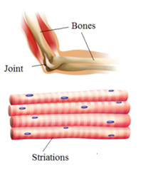
The two ends of skeletal muscles are typically attached to two different bones by a tendon. The joint between the two bones act as a fulcrum and the contraction of the skeletal muscle bring the bone near to other. They are primarily involved in locomotory actions and changes of body postures.
Characteristics of Skeletal or Striated Muscles:
- They are found in limbs
- They are long and cylindrical
- They occur in a bundle
- They are multinucleate
- The sarcolemma is present in them
- Myofibrils are distinct and alternately light and dark striped. Hence called striated muscles.
- The intercalated (between the layer) disks are absent.
- They contract faster and get fatigued soon.
- They are voluntary.
- Innervated (connected with nerves) to the central nervous system.
Visceral Muscles or Smooth Muscles or Nonstriated Muscles:
Visceral muscles are located in the inner walls of hollow visceral organs of the body like the alimentary canal, reproductive tract, etc. They do not exhibit any striation and are smooth in appearance. Hence, they are called smooth muscles (nonstriated muscle).
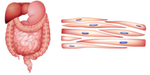
Their activities are not under the control of the nervous system and are therefore known as involuntary muscles. They assist, for example, in the transportation of food through the digestive tract and gametes through the genital tract.
Characteristics of Visceral Muscles:
- They are found in the inner walls of hollow visceral organs of the body like the alimentary canal, reproductive tract, etc.
- They are long and spindle-shaped
- They form a sheet or tube or sphincter.
- They are uninucleate
- Sarcolemma is not present in them and they are bounded by a thin plasma membrane.
- Myofibrils are unstripped and indistinct. Hence called nonstriated muscles.
- The intercalated (between the layer) disks are absent.
- They contract slowly and do not get fatigued. Hence can remain contracted for a long time.
- They are involuntary.
- Innervated (connected with nerves) to the autonomous nervous system.
Cardiac Muscles:
Cardiac muscles are the muscles of the heart. They are striped muscles. Many cardiac muscle cells assemble in a branching pattern to form a cardiac muscle. They are involuntary in nature as the nervous system does not control their activities directly.

Characteristics of Cardiac Muscles:
- They are found in the heart
- They are short, cylindrical, and may be branched.
- They form a network
- They are uni or multinucleate
- The sarcolemma is present and is thin, bounded by a thin plasma membrane
- Myofibrils are distinct and alternately light and dark stripped.
- The intercalated (between the layer) disks are present.
- They contract quickly and never stop and never get fatigued.
- They are involuntary.
- Innervated (connected with nerves) to the autonomous nervous system.
Structure of Muscles:
Structure of Skeletal Muscle
The human body has nearly 639 to 650 skeletal muscles. Each organized skeletal muscle in the human body is made of a number of muscle bundles or muscle fascicles (plural: muscle fasciculi) held together by a common collagenous connective tissue layer called fascia or epimysium. Each muscle bundle contains a number of muscle fibres. Each muscle fascicle is covered by a connective tissue called perimysium.
Each muscle fibre or muscle cell is lined by the plasma membrane called sarcolemma enclosing the sarcoplasm. Muscle fibre is a syncitium as the sarcoplasm contains many nuclei. The cytoplasm containing many nuclei is called syncytial sarcoplasm. The nuclei are located on the periphery of sarcoplasm.
The interior of the sarcoplasm is occupied by the tubules of the endoplasmic reticulum or sarcoplasmic reticulum. It is the is the store house of calcium ions. A characteristic feature of the muscle fibre is the presence of a large number of parallelly arranged filaments in the sarcoplasm called myofilaments or myofibrils.
The cytoplasm contains glycogen granules and many mitochondria.
Structure of Miyofibril:
Each myofibril is about 1 – 3 μm in diameter. It has alternate dark and light bands on it throughout the length. Huxley, Murray, and Waber (1974) discovered that this striated appearance is due to the distribution pattern of two important contractile proteins Actin and Myosin. The light bands contain actin and are called I-bands or Isotropic bands or actin myofilament, whereas the dark bands called ‘A’ or Anisotropic bands or myosin myofilament contain myosin. Both the proteins are arranged as rod-like structures, parallel to each other and also to the longitudinal axis of the myofibrils.
In a resting state, the edges of thin filaments on either side of the thick filaments partially overlap
the free ends of the thick filaments leaving the central part of the thick filaments. This central part of thick filament, not overlapped by thin filaments is called the ‘H’ zone.
Light Bands or Isotropic Bands (I-bands)
- These bands are light coloured bands.
- These are formed of thin actin filaments only.
- They appear nonrefractive under polarized light and hence known as isotropic bands or I-bands.
- These bands are bisected by thin dark elastic fibre line at midpoint called z-line or z-band or Krause’s membrane.
- They are firmly attached to Z-discs.
- The part of a myofibril between two adjacent z-discs is called a sarcomere.
- These bands shorten during muscle contraction.
Dark Bands or Anisotropic Bands (A-bands)
- These bands are dark coloured bands.
- These are formed by thicker myosin and actin myofilaments.
- They are doubly refractive and hence known as anisotropic bands or A-bands.
- These bands are bisected by thin paler line at midpoint called Hensen’s line or H-zone. A narrow dark line passes through H-band and called as M-line or M-band or M- membrane.
- They are free at both ends.
- Length of these bands does not change during muscle contraction.