In this article, we are going to study another important component of blood plasma i.e. White blood corpuscles, their types, structures, and functions.
They are colourless, nucleated, amoeboid (irregular shape) and phagocytic cells larger than RBC’s. Their size ranges from 12 μm to 20 μm. They have mitochondria, Endoplasmic reticulum, Golgi complex, ribosomes, and centrioles. They can move through the walls (squeeze through) of blood vessels into the lymphatic system and tissues. This is known as diapedesis. They exhibit phagocytosis.
The total number of WBCs per milliliter of blood is called total leucocyte count (TLC). Their approximate count in blood of a healthy person is about 5000 to 9000 WBC’s per mL.
They are produced in the bone marrow, lymph glands, spleen, and thymus. The formation of WBCs is called leucopoiesis. The worn-out WBCs are destroyed by phagocytosis in blood, liver, spleen, and lymph nodes. Their life span is about 8 hrs to 3 days.
Types of White Blood Corpuscles (Leucocytes):
Depending upon the presence of granules present in the white blood corpuscles are further classified as a) Granulocytes and b) Agranulocytes
Granulocytes:
They contain granules and are irregular. They contain a multilobed (polymorphic) nucleus in their cytoplasm. Hence the granulocytes are also called polymorphs or polymorphonuclear leucocytes.
Granulocytes on basis of staining agents are classified as i) Neutrophils ii) Eosinophils (Acidophils) and iii) Basophils.
Neutrophils:
They are Stained with neutral dyes. They are the most abundant and constitute about 70 % of total WBCs. They are 12 to 12 μm in size. Their nucleus is 3 to 5 lobed. They are phagocytic in function i.e. they engulf microbes. Their granules are fine and inconspicuous (not clearly visible).
Functions:
- They are motile, and phagocytic. They engulf microbes and destroy damaged tissue and bacteria. Thus they act as scavenger.
- They squeeze out of capillary wall at the site of injury typically accompanying inflammation. The process is called diapedesis.
- They self destruct after one burst of activity.
Eosinophils (Acidophils):
They are stained with acidic dyes like eosin. They constitute about 2 to 3 % of total WBCs. Their nucleus is 2 lobed (bilobed). They are less phagocytic in function than neutrophils. Their number increases in allergy, hay fever, asthma, and parasitic infection. A pathological increase in number is called eosinophilia.
Functions:
- Eosinophils are particularly important in fighting parasitic infections. Eosinophils are capable of phagocytosing bacteria
- They contain hydrolytic enzymes and peroxidases that kill large parasites. They cannot engulf parasites but attach to the parasite and secret chemicals which kill the parasite. They engulf bacterial toxins.
- Eosinophils have antihistamine activity and act as antiallergic.
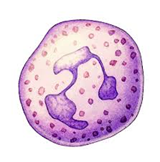
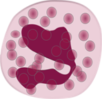
Basophils:
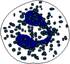
They are Stained with basic dyes like methylene blue. They constitute 0.5 % of total WBCs. Their nucleus is elongated and twisted with two or three lobes. They are non-phagocytic in function. Their granules contain heparin and histamine.
Functions:
- They release heparin (anticoagulant) and histamine involved in inflammatory and allergic reactions. Histamine is released in allergic response and in injured tissues to dilate blood vessels.
Agranulocytes:
Agranulocytes do not contain granules. Agranulocytes are further classified as i) Lymphocytes and ii) Monocytes
Lymphocytes:

These are a smaller size, rounded with a large nucleus. Their size is 6-14 μm. They constitute 20 to 25 % of total WBCs. There are two types of lymphocytes: B-lymphocytes and T-lymphocytes. B-cells develop in the bone marrow. T cells are born in the bone marrow but are matured in the Thymus. They are produced in the bone marrow and mature in spleen and lymph nodes.
Functions:
- They produce antibodies. They are responsible for the immune response of the body.
- The B-cells develop into plasma cells which make antibodies, The T-cells attack viruses, cancer cells, and transplants.
Monocytes:
They are the largest of all WBCs. Their size is 12-20 μm. They have a large kidney-shaped nucleus. They constitute 3 to 7 % of total WBCs. They are called wandering cells of the blood.
Functions:
- They are phagocytic in function. They remove cell debris and act as scavengers of the cell.
- They come out of capillaries into tissues and change into macrophages. Macrophages are monocytes that have migrated out of the bloodstream and into the internal body tissues.
Functions of White Blood Corpuscles:
Phagocytosis:
Phagocytosis is a process by which cells ingest large particles (> 0.5 micrometers) into membrane-bound vesicles called phagosomes, which are then targeted to the lysosomes for enzymatic degradation. On arriving at the site of infection, neutrophils and monocytes engulf microbes or foreign matter or the damaged cell. This process is called phagocytosis. Thus phagocytosis is important for the protection of the body against infection.
Scavenging:
In phagocytosis neutrophils in blood and monocytes in tissues engulf microbes, foreign matter or the damaged cell. Thus they act as a scavenger by removing debris of dead cells and dead microbes from the body.
Pus Formation:
Pus is a whitish substance made of millions of dead WBC’s and living and dead bacteria. Thus pus is formed by exuded plasma, dead WBCs, destroyed tissue cells, living and dead microbes. Pus usually forms when the pathogen is winning the battle.
Diapedesis:
Neutrophils by amoeboid movements squeeze out of the capillary wall at the site of injury and engulf and kill microbes. The process is called diapedesis. It is accompanied by inflammation.
Inflammation or Inflammatory Reaction:
A localized reaction that produces redness, warmth, swelling, and pain as a result of infection, irritation, or injury is called inflammation or inflammatory reaction. Inflammation is a vital part of the body’s immune response. It is the body’s attempt to heal itself and repair damaged tissue.
The blood vessels at the region of injury dilate and release more blood making them hot and red. The tissue fluid at the site of injury accumulates and thus the area swells up.
Neutrophils and monocytes enter the tissues by diapedesis and fight against invading microbes.
Formation of Antibodies:
Antibodies, also known as immunoglobulins, are produced by the immune system to help stop intruders from harming the body. When an intruder (virus, bacteria, foreign material) called antigens enter the body, the immune system comes into action. The immune system creates antibodies to mark the antigen and destroy it. Thus antibodies kill microbes and neutralize toxins produced by them.