The Earth has formed 4.5 billion years ago and on the Earth, which arose the first life form in the form of Prokaryotic cells. These unicellular creatures are primordial and are building blocks of multicellular organisms. Life is supposed to have originated from the oceans which is why embryos of terrestrial and aerial animals still have gill clefts in some phases of their ontogenic development. It took 3 million years for the first cell to have existed on the earth. Prokaryotes cells are extremely simple in their structure. If we split the word ‘PROKARYOT’, we get two words- Pro, meaning Primitive and Karyon, which means the nucleus. The Prokaryotic cells are not as complex as the eukaryotic structures. They did not have a true nucleus and the genetic material was suspended in the cytoplasm called a nucleoid. Example – bacteria.
Structure of Prokaryotic Cell:
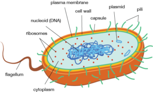
Cell Envelope:
The cell envelope is the outer covering of the cell and gives shape to the cell and protects the cell organelles. It consists of the following 3 layers:
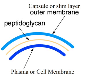
Glycocalyx:
It is found in some of the bacterial cells and is mainly composed of the macromolecules. It protects the contents of the cell and is of two forms: The Capsule and The Slime Layer. The Capsule is thick, strong and provides mechanical support to the cell. It is immunogenic in nature. And because of their thick skin, they sometimes get on people’s nerves. Not literally but this capsule is so strong that it can withstand the attack of the White Blood Cells. Moving on, the capsule is a layer made by the firm gathering of polysaccharides which is a distinguishing character between the capsule and the slime layer. The Slime layer is also known as loose sheath because here the glycoprotein molecules are loosely arranged. This layer helps to maintain moisture in the cell.
Cell Wall:
The cell wall is normally absent in the Prokaryotic cells. However, if it is present, it is made up of poly peptidoglycans. Peptidoglycans are only found in the cell walls of bacteria. It helps in shape maintenance and in osmosis and transporting nutrients in and out of the cells. Peptidoglycans are alternating units of N- acetylglucosamine and N- acetylmuramic acid. They help in the transportation process, for certain nutrients, these bacteria have to use another way. If the nutrients are too large to be taken inside through the pores, certain enzymes are used. These enzymes convert the nutrients into smaller or simpler substances that can be easily taken in by the cell. And the cytoplasm is responsible for the secretion of these ‘Exoenzymes’. However, certain bacteria do not have cell walls. These bacteria use certain proteins as a protective covering. And sometimes this helps in getting the antibiotics. Smart technology there!
Plasma Membrane:
The Plasma Membrane is the innermost covering and is made of amphipathic molecules. That means that these molecules have hydrophilic and hydrophobic ends. Most of these molecules are the proteins, lipids and cholesterols. Universally, The Fluid Mosaic Model has been accepted as a structure of the plasma membrane. This model represents the membrane to be like a sea of lipids with protein icebergs floating on it and in it. The proteins which are completely submerged as known as intrinsic proteins and the ones that are outside the lipid layers are known as the extrinsic proteins. Some protein passes through the lipid layers and they are known as tunnel proteins. Their positions affect their solubility in the lipids. This shall be discussed later. Thus based on the plasma membrane’s mosaic-like structure, Seymour Jonathan Singer and Garth L. Nicolson put forth the FLUID MOSAIC MODEL in the year 1972. The plasma membrane is extremely important for all the life forms. It not only separates the contents but also helps in the exchange of materials and helps in the uptake of the nutrients that are essential to the cell. Numerous activities to take place in the cell membrane.
Organelles:
Prokaryotic cells are primitive cells and hence do not show well-defined membrane-bound organelles like the ones in the eukaryotic cells. But there are some membrane-bound organelles and these are mesosomes and certain pigment containing chromatophores.
Mesosomes:

These are formed by the invaginations of the plasma membrane or the cell membrane. Invaginations are nothing but certain infoldings. These mesosomes are mostly seen in the gram-negative bacteria and can be of the form of tubules, vesicles and lamellae.
They help to form the cell wall and increase its surface area. They especially help in respiration since the respiratory enzymes are associated with them. In eukaryotes, these enzymes are present in the mitochondria. Mesosomes also help in the replication of the DNA. There are two types of mesosomes. Septal: the ones which extend towards the centre of the cell. and Lateral: the ones that are peripheral.
Chromatophores:
The cyanobacteria like Nostoc and Anabaena have chromatophores consisting of the pigments that are necessary for photosynthesis.
Cytoplasm:
The cytoplasm consists of water, enzymes, salts and other compounds. It is like a semi-liquid structure and does not show cytoplasmic streaming or cyclosis. It appears to be granular because of ribosomes and inclusion bodies.
Inclusion Bodies:
- Organic Inclusion Bodies: These are phosphate granules, poly beta-hydroxybutyrate granules, carboxysomes, cyanophycean granules, glycogen, gas vacuoles and more. The gas vacuoles give buoyancy to the aquatic plants. This helps in the process of photosynthesis since the plants can trap sunlight from the atmosphere.
- Inorganic inclusion Bodies: These are Phosphate and sulphur granules. They are known as metachromatic granules due to their ability to take various colours. The phosphate granules store phosphate. The sulphur granules are formed when H2 S is used as a hydrogen donor.
Ribosomes:
In prokaryotic cells, the ribosomes are of a 70S type. These have small 30S subunit and large 50S subunit. The small subunit consists of rRNA of 16s type and the large subunit have 23S and 5S type. Subunits are long RNA with proteins on them. And these subunits help in the protein synthesis by locking in together with each other. The main function of the ribosomes is as mentioned earlier, protein synthesis.
Nucleoid:
As the name prokaryotic suggests, a true nucleus is absent in these kinds of cells. The circular and double-stranded DNA is known as the genome. The histone proteins that are present in the eukaryotic cells are absent in the prokaryotes. The basic function of the histone proteins is to hold the DNA together and it affects the gene regulation. There are 11 types of histone proteins viz. H2A, H2B, H3K4, H3K9, H3K27, H3K36, H4K5, H4K8, H4K12, H4K16, H4K20, each having a different composition. The DNA is 1 micrometre long and is attached to the plasma membrane through the mesosomes. The DNA has 3000-4000 genes. A looped domain can be seen which is a structure formed by the tightly coiled structure of the DNA. This looped domain is held in position by RNA molecules.
Plasmids:
The extrachromosomal, self-replicating units are known as plasmids. The plasmids are circular and have a double-stranded DNA. Plasmids serve as agents for gene transfer and hence are used in Recombinant DNA Technology as vectors or vehicles for carrying proteins. They have anti-biotic, metal and drug resistance. For bacterial fertility, episomes which are a type of plasmid are highly important. Episomes have a self-replication capacity.
Other Structures:
Flagellum, used for locomotion; Pili, used in the mating process. Fimbrae, used for the clinging of the cells and Spinae, the appendages used by the cell for the adjustment to the external environmental conditions like temperature, pH, salinity, etc; are some of the structure present in the Prokaryotic cells.