In this article, we shall study sexual reproduction and the male reproductive system in humans. The process of formation of life from pre-existing life is called reproduction. In animals two types of reproduction are observed a) asexual reproduction and b) sexual reproduction.
Asexual Reproduction:
A mode of reproduction in which the offspring comes from a single organism, and not from the union of gametes as it is in sexual reproduction is called asexual reproduction
Characteristics of asexual reproduction:
- In this mode, an individual can give rise to daughter individuals by mitotic division of a part of its own body. As body cells are involved in the mitotic division. asexual reproduction is also termed as somatogenic reproduction.
- There is the absence of fusion of gametes. Hence asexual reproduction is also called agamogenesis or agamogeny.
- The offspring are genetically identical to the parent and dis exactly same without any variations. Hence the offspring is the clone of parents. Variations can take place only due to mutation.
- It is a faster method of multiplication.
- It may take place by binary fission, budding, vegetative propagation, spore formation (sporogenesis), Fragmentation (Regeneration), Parthenogenesis, apomixis and nucellar embryony.
- It is commonly observed in lower organisms like protists, sponges, coelenterates and certain flatworms.
Sexual Reproduction:
Sexual reproduction is a mode of reproduction that involves fusion of female gamete (ovum) and male gamete (sperms) during fertilization.
Characteristics of sexual reproduction:
- In this method, two individuals are involved in the reproduction.
- There is a fusion of female gamete (ovum) and male gamete (sperms) during fertilization. The result of which zygote is formed, which develops into offspring.
- Due to the fusion of gametes, this mode of reproduction is also termed as gamogenesis or gamogeny.
- As characters of offspring are derived from two different individuals, variations can be observed.
- It is observed in higher organisms.
Human Reproductive System:
Puberty means the changes that occur in boys and girls as they grow up. In this period the maturity of human sex organs begins. Most of these changes occur between the ages of 10 to 14 years. These changes are brought about by certain hormones. During puberty, the body grows rapidly, and both primary and secondary reproductive organs grow and become mature. Along with these changes, secondary sex characters also start appearing.
In males, primary sex organs are male gonads also known as testis. testis produce male gametes (male sex cells) also called sperms or spermatozoa. In females, primary sex organs are the ovary. Ovary produces an ovum, plural: ova (female sex cells) also referred as eggs. The secondary sex organs are different in males and females. They include reproductive ducts for transporting gametes and accessory gland which help reproduction.
In males, sexual maturity is attained at the age of 13–14 years and in females, at the age of 11–13 years. Puberty ultimately leads to a stage when the child becomes an adolescent. The World Health Organization (WHO) defines adolescence as the period from 10 to 19 years of age characterized by developments and changes in physical, psychological, and social areas.
During adolescence, the secondary sexual characters that develop are as follows:
- In males: deepening of the voice, widening of shoulders, muscular body, the appearance of beard and moustache, the growth of axillary and pubic hair, enlargement of external genital organs.
- In females: the growth of axillary and pubic hair, widening of pelvis and hip, enlargement of breasts (mammary glands) and initiation of the menstrual cycle.
The Stages of Human Reproduction:
Human shows sexual reproduction and the changes in the body takes place for viviparity. viviparity means the development of the embryo inside the body of the parent, eventually leading to live birth,
- Formation of gametes (Gametogenesis)
- Changes in the female body to facilitate the entry of sperms during copulation.
- Fusion of gametes. (Fertilization).
- Development of Zygote. (Embryology).
- Production of milk for the nourishment of young ones.
- Hormonal coordination by pituitary glands and gonads.
Human Male Reproductive System:
The male reproductive system consists of parts for production of gametes and copulation.
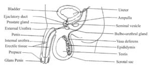
The male reproductive system consists of a pair of testes, a pair of the epididymis, a pair of vasa deferentia (singular: vas deferens), urethra, penis and accessory glands. Teses are sex glands. vasa efferentia, epididymis, ductus deferens and ejaculatory ducts are part of the duct system. Seminal vesicles, prostate and Cowper’s gland form system of accessory glands. The penis is copulatory organ.
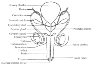
Testes:
They are the male gonads. During early foetal life, the testes develop in the lumbar region of the abdominal cavity just below the kidney. During the seventh month of development, then migrate into the scrotum permanently. Testes descend into the scrotum along with peritoneum, blood vessels and vas deferens. Failure of testes to descend from the abdomen into the scrotum leads to sterility called cryptorchidism.
Testes are extra-abdominal (present outside the abdomen) in a pouch made up of skin and connective tissue called scrotal sac or scrotum that hangs in the region between the legs. The walls of scrotum consist of smooth muscles called dartos tunic muscles. The scrotum is divided into two compartments by muscle septum. Each compartment encloses a testis, epididymis and a testicular end of a spermatic cord.
Scrotum protects the testes and acts as a thermoregulator. A scrotum has little or no fatty insulation hence it keeps the temperature of testes cooler than the body temperature. Thus scrotum helps in maintaining the temperature of testes at about 2-3°C lower than the body temperature. This temperature is suitable for the development of sperms. The temperature of the scrotum is maintained by involuntary muscles that connect the scrotum to the body. The contraction and relaxation of these involuntary muscles move the scrotum near or away from the body when the temperature of surroundings is low or high.
Note: In some seasonally breeding mammals the testes descend into the scrotum in the breeding season and ascends back in the abdomen in the non-breeding state.

Testes are soft, smooth, pinkish oval organs. In an adult male, each testis is approximately 4-5 cm long and about 12 g in weight. They are mesodermal in origin and located outside the abdomen scrotum. They are suspended in the scrotum by the spermatic cord. Each testis is connected to the wall of the scrotum by a short fibromuscular band called gubernaculum.
Histology of Testis:
The testis is externally covered by fibrous connective tissue called tunica albuginea. It is covered internally by tunica vascularis formed by capillaries and externally by an incomplete peritoneal covering called tunica vaginalis.
Transverse section of testis shows different stages of spermatogenesis like spermatogonia, primary and secondary spermatocytes, spermatids and sperms.
In each testis, there are 200 to 300 lobules. In each lobule, there are 1 to 4 convoluted loops called seminiferous tubules. Each tubule is 70-80 cm length when stretched out. the basement membrane of the seminiferous tubule is lined with highly specialized cells called spermatogonia. The spermatozoa occupy the central part of the lumen of the seminiferous tubule. The cells known as cells of Sertoli are also attached to the basement membrane. These cells provide mechanical support and protection to developing sperms and also participate in their nutrition and maturation.
In between seminiferous tubules masses of cells called interstitial cells or Leydig cells are present. These cells secrete the male hormone, testosterone, which is responsible for development and maintenance of male sex characteristics. The interstitial connective tissue also contains fibroblasts, blood vessels, nerves and lymphatics.
seminiferous tubules converge at the posterior surface and form a network of irregular tubules called rete testis.
Epididymis:
These are a pair of ‘C’ shaped structures each lying along the posterior border of each testis. It is a long (6 m) highly coiled tube which remains attached to the testis and lies within the scrotal sac. The long length of the epididymis delays the release of the sperm and allows them time to mature. Epididymis stores spermatozoa (sperms) and serves as a passage for their transport from the testis.
The epididymis consists of three parts: head (caput epididymis), body (corpus epididymis), and tail (cauda epididymis). The head of the epididymis is located on superior pole of the testis. It stores sperm for maturation. Here sperms acquire increased motility and fertilizing capacity. The tail stores sperms for short period before they enter vas deferens. The tail of the epididymis is continuous with the vas deferens.
Vas Deferens (Sperm Duct):
Each cauda epididymis continues as vas deferens. Each vas deferens is about 40 cm long and enters the abdominal cavity, passes
over the urinary bladder and joins the duct of seminal vesicle to form the ejaculatory duct.
Ejaculatory Duct:
These are the pair of ducts each about 2 cm long. It is formed by joining of vas deferens and a duct of the seminal vesicle. Both the ejaculatory ducts open into the urethra in the region of the prostate gland. They carry seminal fluid and spermatozoa to the urethra.
Urethra:
The urethra in males is about 15-20 cm long and is differentiated into three parts an anterior prostatic part which passes through the prostate gland; a middle membranous part; and a posterior penile part which passes through the copulatory organ, the penis. Urethra functions as a passage for both semen and urine.
The urethra is the tube that carries urine from the bladder to outside of the body. In males, it has the additional function of expelling (ejaculating) semen when the man reaches orgasm. When the penis is erect during sex, the flow of urine is blocked from the urethra, allowing only semen to be ejaculated at orgasm.
Penis:
The penis is a cylindrical, spongy, muscular, a highly vascular (supplied with blood vessels), erectile and pendulous copulatory organ in males. It is suspended in pubic region in front of the scrotum. The urethra runs through it centrally throughout its length. It contains three columns of erectile tissues. Ordinarily, it remains small and limp. During sexual excitement, the spongy tissue gets filled-up with blood, making it erect, long and stiff.
Externally, the penis is covered by skin. Near the tip of penis, corpus spongiosum is enlarged making it soft and highly sensitive. It is called glans penis. It is covered by a loose fold of skin called prepuce or foreskin which can be retracted.
The penis contains two posterolateral tissues called corpora cavernosa and median corpus spongiosum. Urethra passes through spongiosum, hence it is called spongial urethra.
Accessory Sex Glands of Male Reproductive System:
Seminal Vescicles:
A pair of seminal vesicles is present at the base and posterior side of the urinary bladder. They are fibromuscular pouches. The seminal vesicles store sperms that descend from the testis and secrete seminal fluid. The seminal fluid is a viscous fluid and contains fructose, fibrinogen and prostaglandins. Fructose provides energy to sperms for swimming. The prostaglandins stimulate contraction in the female reproductive tract to help in the process of fertilization. The fibrinogen helps in coagulation of semen after ejaculation. Seminal fluid forms about 40-80 percent of the ejaculate (semen thrown out of the penis).
Prostate Gland:
Prostrate gland surrounds the first part of the urethra. It consists of 20 to 30 separate lobes which open separately into the urethra. It secretes an alkaline fluid which is discharged into the urethra. This fluid keeps the sperms alive and helps them to swim vigorously.
Due to its alkaline nature, it neutralizes the acidity of vaginal secretion and helps in maintaining the pH at 6.0 to 6.5. At this pH the sperms become motile and facilitate the process of fertilization. Secretion of prostate gland forms about 5-30 percent of the ejaculate.
Cowper’s Glands or Bulbo-Urethral Glands:
These are paired glands that lie below the prostate gland and join the urethra at a short distance from that of the prostate gland. They are pea-sized and situated on either side of the membranous urethra. Cowper’s glands secrete a white, viscous, alkaline secretion resembling mucus. It neutralizes acids that may be present in the penile urethra due to previous urination. It also lubricates vagina of the female genital tract.
Spermatozoa and Semen:
The semen is ejaculated during sexual intercourse called coitus. Semen is a whitish fluid containing spermatozoa and a mixture of secretions from seminal vesicles, prostate glands and Cowper’s gland. The process of expulsion of semen from the urethra is called ejaculation.
The process of formation of sperms or spermatozoa is termed Spermatogenesis. The spermatozoa are male gametes produced by the testes. Human sperm has three main parts head, neck and tail. The tip of a sperm is covered by
a cap-like structure, acrosome, which helps the sperm to penetrate inside the egg during fertilization.
In epididymis, spermatozoa are stored and they are non-motile. By the secretions from the accessory reproductive glands in males, they get activated and motile.
The sperms are released in millions. In one ejaculation 3 to 4 ml of semen is produced containing about 300,000,000 (3 × 10⁸) sperms are discharged but only one of the fertilizes the egg. The release of a large number of sperms ensures the process of fertilization.
Sperms when introduced into the vagina of the female move with the speed of 2 mm/minute inside the body of the female.